3dPrintedOrgans
Latest
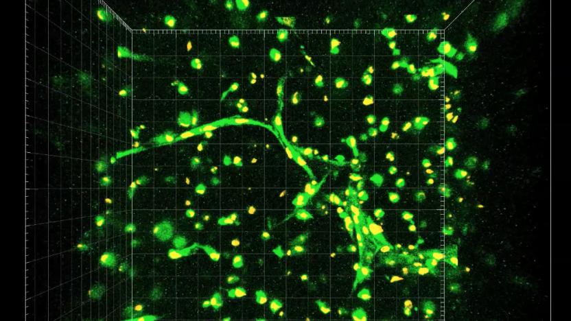
Researchers are closer to working capillaries in 3D-printed organs
Researchers have brought 3D-printed organ and tissue capabilities a long way, but the technology still faces a few challenges. A major one is how to incorporate blood vessels into bioprinted tissue. Living tissue needs a blood supply nearby because without blood to bring in nutrients and take away waste, biological cells will die. Researchers have been able to print larger blood vessels, but functional small vessels like capillaries have been much harder to create and sustain. However, researchers at Rice University and Baylor College of Medicine have developed a new technique, published in Biomaterials Science, that might make it possible.

Doctors reveal they can 3D print body parts and tissue
3D printing isn't just for toys and models -- doctors at the Wake Forest Institute for Regenerative Medicine announced yesterday that they've managed to 3D print "living" tissue and organs that functioned properly when implanted in animals. The team, led by Anthony Atala, is already renowned for printing the building blocks for human bladders. But now they've reached another level entirely: They say it's possible to print structures large and strong enough for humans. They've printed muscle structures, bone and ear tissue so far, according to Nature. With a little more work, the technology could revolutionize the way we approach surgical replacements (and finally make plenty of sci-fi biotechnology scenarios a reality).

Boston Children's Hospital preps surgeons with custom 3D-printed models
Undoubtedly, 3D printing has taken root in a variety of disciplines, and medicine is no stranger to leveraging its tool kit. At Boston Children's Hospital, surgeons are using printed models to prep for the operating room. "With 3D printing, we're taking a step that allows experienced doctors to simulate the specific anatomy of their patients and allows the best of the best to become even better," says Peter Weinstock, MD, PhD. Dr. Weinstock is working on an in-house service that's capable of constructing the models in short order. Using scans from the hospital's radiology department and a 3D printer capable of super high-resolution output (16 microns, to be exact), the models allow doctors to examine details of a baby's skull or brain. What's more, the machine can use multiple materials to sculpt the final result, simulating the unique facets of bone, skin and blood vessels individually. For surgeons-in-training, the custom-made prints can illustrate the details of a medical condition rather than an average look.

Researchers use 3D printer, sugar, to create a fake artery network for lab-grown tissue
Printing a chocolate heart is easy enough, but how about an actual organ? There are folks working on it, but it turns out those veins of yours aren't exactly a breeze to replicate. Researchers at the University of Pennsylvania and MIT may have found a semi-sweet solution -- dissolving a sugar lattice in a batch of living Jell-O. The research team uses a RepRap 3D printer and a custom extruder head to print a filament network composed of sucrose, glucose and dextran which is later encased in a bio-gel containing living cells. Once the confectionery paths are dissolved, they leave a network of artery-like channels in their void. Tissue living in the gel can then receive oxygen and nutrients through the hollow pipes. The research has been promising so far, and has increased the number of functional liver cells the team has been able to maintain in artificial tissues. These results suggest the technique could have future research possibilities in developing lab-grown organs. MIT Professor Sangeeta Bhatia, who helped conduct the effort, hopes to push the group's work further. "More work will be needed to learn how to directly connect these types of vascular networks to natural blood vessels while at the same time investigating fundamental interactions between the liver cells and the patterned vasculature. It's an exciting future ahead." Scientists at other labs could also get their mitts on the sweet templates since they're stable enough to endure shipping. Head past the break for a video of the innard infrastructure.


