microscopy
Latest
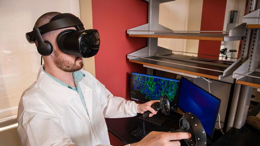
VR and microscopy help scientists see 'inside' diseases
You can only learn so much about cells by studying 2D pictures, and 3D microscope technology can produce an abundance of data that might be hard to decipher. Researchers at Carnegie Mellon University and Virginia Mason have an answer, though: let scientists walk 'inside' the cells. They've combined virtual reality with expansion microscopy (which grows samples by over 100 times) to explore cell data that would otherwise be too complex to handle. Once the cells have been imaged, labeled and compiled into data, a custom technique turns the 2D info into 3D environments.

Researchers use electric currents to detect cancer in human tissue
In a study published recently in Angewandte Chemie, researchers demonstrated that an imaging technique called scanning electrochemical microscopy could become a very useful medical tool. Rather than having to use additional chemicals like dyes or fluorescent markers to get a good look at tissue, this method uses electrochemical probes to detect natural biomolecules around the tissue.

Extremely detailed images of living cells can now be taken over time
Advanced microscope technology now lets us view objects at the nanoscale, meaning, when it comes to biology, we can see details of living cells that were never possible before. But doing that comes with a few requirements that have been fairly limiting. For instance, you have to be able to pack a lot of fluorescent dye into the object you want to see and you need that dye to be really stable. Typically, those sorts of dyes grab onto proteins in the object, but proteins are often not distributed densely enough, limiting how much dye can be introduced. Also, these fluorescent dyes tend to bleach out really quickly, only giving researchers a few seconds of imaging time.
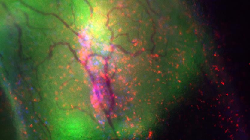
Scientists watch an immune system fight the flu in real time
To date, biologists have typically had to study the progress of a virus through indirect means, such as studying the antibodies -- actually tracking the viruses themselves has been difficult. However, researchers say they've found a way to follow the progress of a virus in real time. By using multiphoton microscopy in tandem with a laser and fluorescence, the team monitored influenza virus in a mouse's trachea (where the transluency made imaging possible) through the infection and immune system response.

Scientists model a Coronavirus' infectious bits for the first time
A collaboration of scientists from University of Washington (UW), the Pasteur Institute and the University of Utrecht have harnessed a state-of-the-art microscope and supercomputer to model a coronavirus' infection mechanism for the first time.

Scientist trumps his own work three weeks after winning the Nobel Prize
If you'd just won the Nobel Prize for Chemistry, no-one would blame you if you took a quick trip to Disneyland, or at least a few days to catch up on Orange is the New Black. Eric Betzig, however, had other plans, since shortly after he was told he was receiving the accolade for revolutionizing the world of microscopy, he was ready to do it all over again. Put (very) simply, his first achievement, PALM, was a microscope capable of observing cellular interactions in unprecedented detail. The downside to the technique, however, was that it couldn't take shots of fast-moving cells, produced images with a halo around them and the light used to take the pictures was toxic to the cells being studied.

So you want to turn your iPhone into an underwater microscope...
Have you ever wanted to turn your iPhone into an underwater microscope? Of course you have! And starting today, the folks at Bodelin Technologies -- the same folks who brought you the ProScope Micro Mobile that we reviewed last year -- have a way that you can go scuba diving and take an illuminated microscope with you. Basically, they've married the US$149 ProScope Micro Mobile to the UK-manufactured Aquapac, making it very easy for you to use the powerful iPhone microscope underwater. You don't need to be a scuba diver, though -- the video below shows the microscope being used in a fish tank and also in some shallow water on a beach. As Bodelin's Peter White points out, allowing microscopy in the field has some great benefits. Says White, "For marine biology, this is even more important as you won't be removing a plant or animal from their natural environment to examine them under magnification. For corrosion testing, an engineer can closely examine a pipeline, bridge structure or vessel without having to remove a sample to send to the surface." The Aquapac 418 used with the ProScope can be purchased from Bodelin for just $40.

Researchers turn standard microscope into billion-pixel imaging beast
A team of researchers at the California Institute of Technology, led by Professor Changhuei Yang, have figured out a way to crank their microscopy up to 11. Usually, scientists are forced between a rock and a hard place: they can have high res images of small areas or low resolution pictures of larger fields. Using a strategy known as Fourier ptychographic microscopy, Yang's team was able to computationally correct a standard microscope's low res imagery, producing a billion-pixel picture. By adding an LED array to an existing microscope -- the only hardware tweak their $200 system calls for -- the researchers were able to stitch together a 20X quality image from a 2X optical lens. The information gleaned from the LED lights was corrected entirely on a computer, making it an exceptionally cost effective way to create high res microscopic images. The team's report, published by the journal Nature Phototonics, can be read in full at the source link below.

Scientists generate 281-gigapixel cell map using electron microscope
Electron microscopes can produce incredibly detailed and even 3D views of sub-cellular structures, but often at the cost of losing the bigger picture. Researchers at Leiden University in the Netherlands, however, have leveraged a technique called virtual nanoscopy that enables researchers to observe the whole of a cell and its intricate details in a single image. With the method, the team stitches together nanometer resolution photographs of what's gone under the scope to create a map with adjustable zoom a la Google Maps. Their study created a 281-gigapixel image (packed with 16 million pixels per inch) of a 1.5-millimeter-long zebrafish embryo. If you'd like to take a gander at the ultra-high resolution fish or read up on the group's findings for yourself, check out the source links below.

New high-res imaging could make biopsies obsolete, doctors still cutting up in meantime
So maybe a true-to-life Innerspace is still a few years off, but a professor at the University of Rochester has developed a way to take high-resolution 3D images under the skin's surface, potentially eliminating the need for biopsies in cancer detection. Professor Jannick Rolland created a prototype that uses a liquid lens, in which a droplet of water replaces the standard glass lens, in conjunction with near-infrared light, to take thousands of pictures at varying depths. Those images are then combined to create clear, 3D renderings of what lies up to one millimeter below your epidermis. The method has already been tested on livings beings, but is likely a long way from making it to your doctor's office, which means it's off to the guillotine for that Pangaea-shaped mole you've been picking at.

World's smallest periscope provides multi-dimensional view of cells
We never thought we'd say this, but the standard microscope's day may be coming to an end. Okay, so maybe that's a stretch, but a new device conjured up by scientists at Vanderbilt University sure could stand in as a suitable and deserving replacement. In what's being described as the world's smallest version of the periscope, the so-called mirrored pyramidal wells are being used to allow researchers to see several sides of cells simultaneously. The pyramidal-shaped cavities are molded into silicon "whose interior surfaces are coated with a reflective layer of gold or platinum," and when a cell is placed inside, it gives Earthlings a magical multi-dimensional view. It's said that this technology is actually stupendously inexpensive compared to other methods of 3D microscopy, and according to Vandy's own Ron Reiserer, this "could easily become as ubiquitous as the microscope slide." Them's fightin' words, no?[Via Physorg]

NanoPass needles set to vaccinate sans pain
Given the choice, even we'd take the pills over the vaccination, but a new Israeli startup is hoping to ease the fears so commonly associated with needles. NanoPass Technologies is working to develop its "proprietary intradermal drug delivery technology," which supposedly deliver injections without the painful side effects by actually not reaching the nerve endings of the skin. Based on MicroPyramids, which are manufactured by MEMS (Micro Electro Mechanical Systems), the pure silicone crystals are used in extremely diminutive microneedles for intradermal injections, and the tip of the device measures less than one-micrometer in diameter. The company touts its pain-free technology (sound familiar?) as a breakthrough that is "non-intimitdating," which should reduce the likelihood of fainting both youngsters (okay, and adults) face when dealing with needles, and is even said to be easier to administer. Unfortunately, we've got no good news proclaiming that these will be replacing intramuscular and subcutaneous methods later this week, but the $6.5 million in funding that the company has acquired should go pretty far is helping its cause. [Via MedGadget]


