MIT's AI can identify breast cancer risk as reliably as a radiologist
The deep-learning model accurately assesses dense breast tissue.
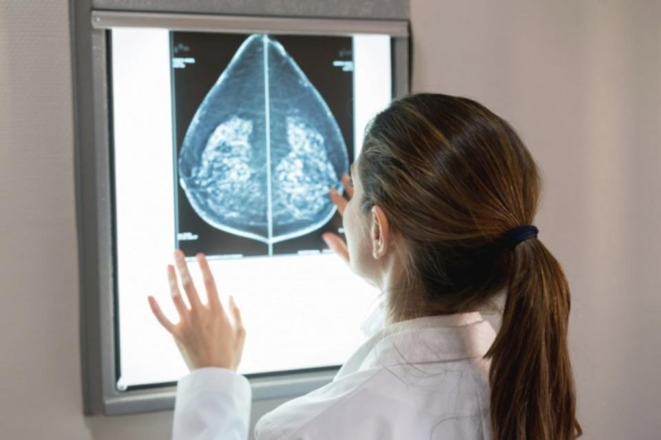

Breast cancer affects one in eight women in the US. There are multiple factors involved in developing the disease, but one issue is dense breast tissue. Some 40 percent of US women have dense breast tissue, which alone increases the risk of breast cancer, and can make mammogram screening more difficult. Now, researchers from MIT and Massachusetts General Hospital (MGH) have developed an automated model that assesses dense breast tissue in mammograms as reliably as expert radiologists.
Breast density assessments have traditionally relied on subjective human exams and calculations, but the deep-learning model -- trained on tens of thousands of digital mammograms -- is able to distinguish different types of breast tissue, from fatty to extremely dense, with 90 percent correlation to radiologists' diagnosis.
In comparison to traditional prediction models, the researchers used a metric called a kappa score, where 1 indicates the model and human experts agree on a diagnosis every time, and anything lower indicates fewer instances of agreements. The maximum kappa score for existing automatic density assessment models is around 0.6. In the clinical application, the new model scored 0.85, meaning it makes better predictions than previous systems.

"Breast density is an independent risk factor that drives how we communicate with women about their cancer risk. Our motivation was to create an accurate and consistent tool, that can be shared and used across health care systems," says MIT PhD student and second author on the model's paper, Adam Yala. "It takes less than a second per image ... [and can be] easily and cheaply scaled throughout hospitals." The researchers now plan on exploring how the algorithm can be transitioned into other hospitals, and how it can be used in other healthcare applications.
