ElectronMicroscope
Latest
Scientists capture 4D atomic movement in breakthrough experiment
Long-held theories about how materials melt, freeze and evaporate may need to be tweaked thanks to some breakthrough research. A UCLA-led team of scientists have captured the 4D movement of atoms through time and 3D space as they changed states, reportedly for the first time. The results were surprising and contradicted classical theories about "nucleation," when atoms start to change from one form to another. The research may prove valuable for the creation and study of new materials, chemicals and biological processes.
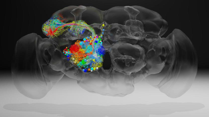
Researchers capture high-resolution image of a complete fruit fly brain
Scientists have created a high-resolution image of a fruit fly brain that will let researchers trace the connections of neurons throughout the brain. A team at the Howard Hughes Medical Institute's Janelia Research Campus led the work, which was recently published in Cell. Davi Bock, the lead researcher on the project, said in a statement that this level of resolution hasn't been achieved before and it will allow scientists to better understand which neurons play a role in behaviors exhibited by fruit flies.
High-powered microscope can scan cells without destroying them
Confocal microscopes are pretty wild. The instruments can capture cell division in realtime, but the downside is the lasers in existing ones tend to fry the cells they're studying. Researchers from the Hong Kong University of Science and Technology may have found a way around that, though. Phys.org explains that professors Du Shenwang and Michael Loy have developed a microscope that's "1,000 times less photo-toxic" than what's available currently.
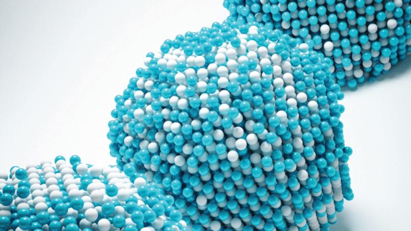
Scientists map every atom inside a nanoparticle
Even the smallest defects can create serious problems. It's a good thing, then, that researchers have found a way to map nanoparticles at an "unprecedented" level of detail -- they've located the 3D positions of all 23,000 atoms in an iron-platinum particle. The group used an extremely high-resolution transmission electron microscope (TEAM I) to capture 2D projections of the nanoparticle's structure, and used an algorithm to stitch those together into a 3D reconstruction. If there's a missing or misplaced atom, you could easily spot it.

This is the world's smallest and happiest snowman
The holiday season gets bigger and more hectic every year -- and maybe you're looking for a smaller, more adorable way to celebrate. Here's one: the world's smallest snowman. This microscopic frosty was built by the nanofabrication lab at London's Western University. He's adorable, but you'll need an electron microscope to see him in person: according to the lab's Tumblr page, the snowman stands at just three microns tall, or about 0.003mm. For comparison, a human hair is about 75 microns thick (0.075mm).

Scientists find the first known dinosaur brain tissue fossil
That lump you see above many not look like much at first blush, but it's a big deal for paleontology: scientists say they have discovered that the sample has the first known example of a dinosaur brain tissue fossil. The team used a scanning electron microscope to detect mineralized blood vessels, collagen, membranes and possibly brain cortex in the remains of an iguanodonid that lived about 133 million years ago. The findings suggest that the dino's brain had a lot in common with those of modern birds and reptiles. Instead of completely filling the cranial cavity, the brain matter significant space for blood vessels and sinuses.

Electron microscope draws nano-sized patterns in metal ink
One of the greatest challenges in designing electronics is drawing very fine details. You normally need lithography, which complicates the process by requiring masks. However, Oak Ridge National Laboratory has now found a way to write at an extremely fine level -- and even get a little bit creative. Its researchers have developed a technique that relies on an electron microscope to draw nanoscale patterns using metal ink. The team first creates a grayscale template to guide its work, and uses the microscope to shoot electrons into palladium chloride cells along that template. The cells neatly deposit raw palladium wherever the microscope goes.

Scientists take detailed pictures of the smallest known life forms
Just how small can life get? Almost unbelievably small, if you ask a team of Berkeley Lab researchers. They've taken the first detailed electron microscope pictures of the tiniest bacteria known to date -- at a typical 0.009 cubic microns in volume, you could fit 150 of them in an already miniscule e. coli cell. Scientists had to catch the hard-to-spot microbes by using a new portable cryo plunger, which flash-froze groundwater to near absolute zero (about -458F) to keep the cells intact while they were in transit.

Electron microscopes stop thieves from covering their tracks
Ask the police and they'll tell you that serial numbers seldom help catch thieves -- dedicated crooks are usually smart enough to file off those digits so that stolen items can't be linked to a crime. Researchers at the National Institute of Standards and Technology might have just found a way to recover those numbers and stop criminals in their tracks, however. Their new technique uses electron microscopes to spot damaged crystal patterns in steel, revealing characters even when they've been polished into oblivion. Current recovery approaches (like acid etching or electrolytic polishing) only sometimes work, and frequently provide faint clues at best -- the microscope produces clear evidence that you could use to convict someone in court.

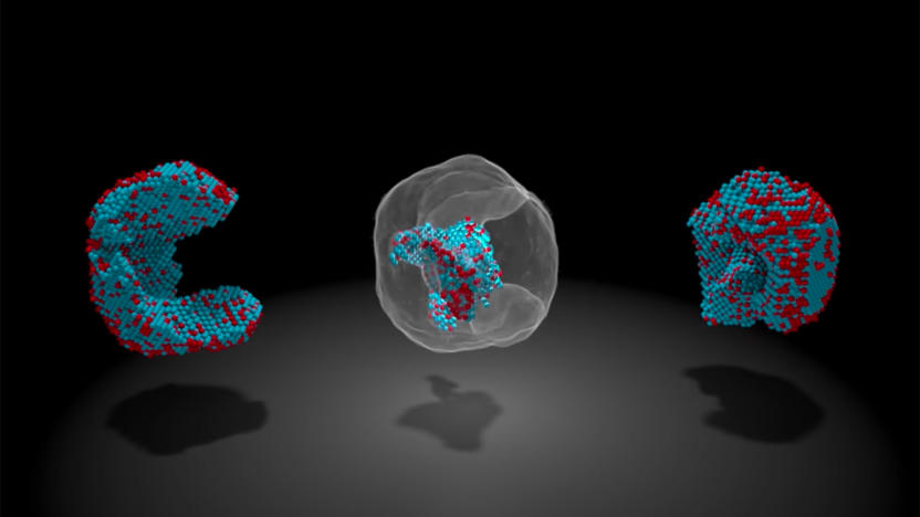
This is what brain synapses look like in 3D
Many know that brains are inherently complex things; there are trillions of synapses converting chemical and electrical signals in a human mind. However, did you know that even those synapses are very complex? If not, it should be perfectly clear now. German scientists have used a mix of extremely high-resolution microscopes (both electron and fluorescent), mass spectrometry and protein detection to create a super-detailed 3D map of a synapse in a rat's brain. It's almost like a miniscule city -- those dots you see represent 300,000 proteins, and only a tiny portion (the glowing red patch at the bottom) is transmitting chemicals.

Electron showers could create the nano-spacesuit of the future
Historically, whenever man or beast's been bombarded with massive amounts of radiation the results have either been gruesome or wholly fantastical (see: any superhero origin story). But recent research out of Japan indicates that a barrage of electrons could actually help scientists revolutionize microbiology and, more excitingly, space travel. The experiment, conducted by a team from the Hamamatsu University of Medicine, found that the larvae of fruit flies hit with this electron rush were able to withstand an electron microscope's hostile vacuum unharmed and even grew to be healthy adults. The results weren't so rosy for the untreated group which, understandably, suffered a grislier fate: death by dehydration. The magic, it turns out, is in that subatomic spray, as the group treated with an electron shower benefited from a polymerizing effect or, more plainly, a bonding of molecules just above the skin's surface that yielded a tough, protective nano-layer measuring between 50- to 100-billionths of a meter thick. Finesse that technique some and it's easy to why one NASA scientist thinks this could lead to the creation of a super-thin "space shield... that could protect against dehydration and radiation." The process is still far from foolproof, however, seeing as how an increase in the microscope's resolution requires an equal boost in radiation -- all of which is fatal to the insects. So, in order to go deeper and get a more close-up view of the larvae's internals, the team's currently exploring new methods of fabricating these "nano-suits" using an array of chemicals. If you're wondering just how far-off we are from practical human application, then consider this: the amount of radiation required to form the bonded layer is akin to "sunbathing naked on the top of Everest under a hole in the ozone." Which is to say, keep dreaming. And get Jeff Goldblum on the phone while you're at it... we have a promising idea for a Return of the Fly sequel.

Scientists generate 281-gigapixel cell map using electron microscope
Electron microscopes can produce incredibly detailed and even 3D views of sub-cellular structures, but often at the cost of losing the bigger picture. Researchers at Leiden University in the Netherlands, however, have leveraged a technique called virtual nanoscopy that enables researchers to observe the whole of a cell and its intricate details in a single image. With the method, the team stitches together nanometer resolution photographs of what's gone under the scope to create a map with adjustable zoom a la Google Maps. Their study created a 281-gigapixel image (packed with 16 million pixels per inch) of a 1.5-millimeter-long zebrafish embryo. If you'd like to take a gander at the ultra-high resolution fish or read up on the group's findings for yourself, check out the source links below.

Beam-switching endows electron microscopes with 3D, added gross-out
Having haunted our curtailed childhoods with tiny, disgusting horrors, the scanning electron microscope is about to get a new lease of life in 3D. Researchers in Japan have figured out how to deflect the electron beam rapidly to give two slightly shifted views, so real-time 3D images can now been scoped on a monitor without even the need for eye-wear. Current gear can only muster flat images, so it's always been painfully slow for scientists to extract convexity and other details from objects. Though the 3D-version is lower-res than the old way, at least now all those slimy mandibles and egg sacs will be right there in your face. Nice.

Periodic table blasted onto a single human hair using ions, human reportedly wants his hair back
We've seen the Torah inscribed on a surface the size of a pin, and the atomic pen making inroads into even more impressive feats, but tiny writing never ceases to amaze us. Now, it seems, the entire periodic table of the elements has been scribed onto a single hair -- that of Martyn Poliakoff, Professor of Chemistry at the University of Nottingham. The project involved magnifying the hair under an electron microscope, and 'writing' on it with ions using an ion beam writer to imprint the entire table of elements onto the hair. As you'll see in the video after the break, the results are quite impressive albeit very small.

Nanotube breakthrough creates scalable transistors
Nanotubes have certainly played their part in various forms of swank gadgetry over the years, but researchers at the University of Illinois, Lehigh University, and Purdue University seem to have upped the ante for future nanotube implementations. Their approach utilizes "dense arrays of aligned and linear nanotubes as a thin-film semiconductor material suitable for integration into electronic devices," which essentially means that the arrays can be transferred into devices where silicon isn't entirely comfortable, such as "flexible displays, structural health monitors, and heads-up displays." Interestingly, the creators aren't expecting their discovery to overtake silicon, but they did mention that the linear arrays could be "added to a silicon chip and exploited for particular purposes, such as higher speed operation, higher power capacity, and linear behavior for enhanced functionality." Sounds like these gurus are just the type Intel would be scouting right about now, eh?[Via TechnologyReview]


