ScanningElectronMicroscope
Latest
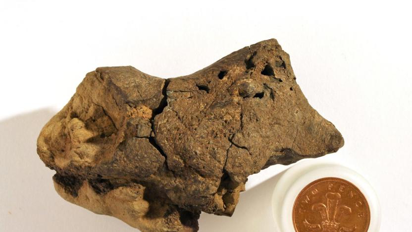
Scientists find the first known dinosaur brain tissue fossil
That lump you see above many not look like much at first blush, but it's a big deal for paleontology: scientists say they have discovered that the sample has the first known example of a dinosaur brain tissue fossil. The team used a scanning electron microscope to detect mineralized blood vessels, collagen, membranes and possibly brain cortex in the remains of an iguanodonid that lived about 133 million years ago. The findings suggest that the dino's brain had a lot in common with those of modern birds and reptiles. Instead of completely filling the cranial cavity, the brain matter significant space for blood vessels and sinuses.

Beam-switching endows electron microscopes with 3D, added gross-out
Having haunted our curtailed childhoods with tiny, disgusting horrors, the scanning electron microscope is about to get a new lease of life in 3D. Researchers in Japan have figured out how to deflect the electron beam rapidly to give two slightly shifted views, so real-time 3D images can now been scoped on a monitor without even the need for eye-wear. Current gear can only muster flat images, so it's always been painfully slow for scientists to extract convexity and other details from objects. Though the 3D-version is lower-res than the old way, at least now all those slimy mandibles and egg sacs will be right there in your face. Nice.

