MRI scans used to create 3D-printed hearts for surgery practice
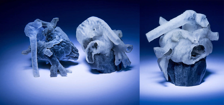

Heart surgeries could be so much safer if surgeons can see and feel an actual representation of the patient's organ before the procedure itself. A system developed by a group of researchers from MIT and Boston Children's Hospital might make that a viable option. By using MRI scans as a blueprint, it allows doctors to print out a model of the patient's heart within just three hours. When you do an MRI scan, the machine takes hundreds of cross-sectional images of your organs. In order to create an accurate 3D-printable model, though, the boundaries between each part of the organ must be determined. It's critical for each part to look distinct, especially if the patient needs surgery due to an unusual anatomy.
If a human manually designates boundaries for each of the heart's parts in the 200-plus scans needed to created a model, it would take him 10 hours. Since that's just too much time, one of the researchers developed an algorithm that speeds up the process. The person in charge of marking boundaries only need to work on around eight cross sections, after which the algorithm will take over to finish the job in an hour. The printing itself takes just a couple of hours more.
Note that the technology's still young, and seven cardiac surgeons at the Boston Children's Hospital will still have to assess how useful the 3D-printed models are this fall. Just recently, select surgeons from the facility started practicing procedures on 3D-printed vasculature. They found that the preparation cut some surgeries' time considerably, making them a lot safer for the patients.
[Image credit: Bryce Vickmark]