Scientists map world first 3D image of cancer-spreading protein
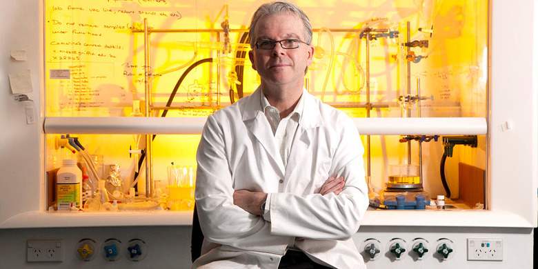
Scientists at Griffith University's Institute for Glycomics released the world's first 3-D image of a protein that is linked to the spread of cancer. Where before scientists had to guess what the structure looked like, now they have a clear 3-D model meaning they can see how it works and develop targeted medicines to stop it before the protein can make matters worse. It was mapped using a technique called X-Ray crystallography and the team, lead by the institute's Director Professor Mark von Itzstein, notes that the image is so well defined that it shows both the structure of the overall protein as well as atomic-level details.
The 3D image is of a bacterial heparanase, which is an enzyme that can break down certain sugars. The human protein is functionally identical to the bacterial one and is found to be over-expressed in cancers. It's also known to play a part in a process called angiogenesis (creating new blood vessels from pre-existing ones) as well as inflammation and the eventual spread of cancer. Because of what this protein does, it is an excellent target for synthesized drugs.
"We explored this [protein] by mutating certain amino acids that kill the activity so that we can understand how the enzyme works," said Professor von Itzstein. "This research has been 10 years in the making and we will now turn our focus to developing a novel anti-cancer drug."
[Image credit: Griffith University]