Scientists unveil 3D microscope that visualizes cells without damaging them
It's like a tiny MRI machine for cells.
There's a problem in cell biology research: to study what happens inside a cell, it has to be destroyed. When scientists use a traditional microscope to observe a cell, they use stains — chemicals that color parts of the cell to make them visible. However, these stains cause damage and kill the cell prematurely. This might not be a problem for long though, as scientists at the Swiss Federal Institute of Technology (EPFL) have developed a technique to look inside living cells without damaging them.


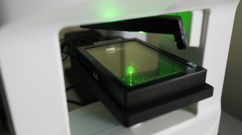
A new 3D microscope, called CX-A, can see the internal structure of cells down to the organelles with a resolution of less than 200nm. The microscope works like an MRI machine for cells, taking images from all angles which can be pieced together into a 3D image. The cell is illuminated by a rotating laser which produces a hologram image of the cell in a non-invasive way.
The automated microscope was unveiled by the Nanolive group and comes with software to convert the readings into 3D images with key cell components shown in color.
This technology will allow scientists to study cells throughout their lifespan. Cells can be observed over periods of hours, days or weeks, so scientists can perform experiments by introducing stimuli and seeing how cells react over time. The user selects how often images should be taken and leaves the machine to run automatically for as long as needed.
This could be used for research into "how biological processes work, how organelles interact and how mitochondria form intricate networks," the EPFL says.