VR and microscopy help scientists see 'inside' diseases
It can show details you wouldn't catch in 2D.
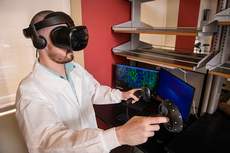
You can only learn so much about cells by studying 2D pictures, and 3D microscope technology can produce an abundance of data that might be hard to decipher. Researchers at Carnegie Mellon University and Virginia Mason have an answer, though: let scientists walk 'inside' the cells. They've combined virtual reality with expansion microscopy (which grows samples by over 100 times) to explore cell data that would otherwise be too complex to handle. Once the cells have been imaged, labeled and compiled into data, a custom technique turns the 2D info into 3D environments.


The approach could be vital to medicine. Ultimately, the team hopes its tool (ExMicroVR) will provide a greater level of understanding about diseases that could lead to more effective treatments. As many as six people can experience data at the same time, ensuring that scientists can work together. And crucially, Carnegie Mellon wants this to be "affordable and easily accessible" to developing countries. You wouldn't need costly microscopes or an abundance of studying time to further medical research.